Teaching Materials
Introductory Biomedical Imaging is targeted at undergraduate students having at least some familiarity with physics and mathematics. The book’s primary content is accessible to students who have taken one year of college physics and one year of college mathematics (ideally calculus). However, the level can be increased by covering material in the otherwise optional “Boxes.” Significantly, no prior knowledge of biology or chemistry is required. This page compiles materials, separated into course materials and textbook materials, that should be useful to be professors teaching a biomedical imaging course and students taking such a course.
Course Materials
This section compiles materials from a one-semester course entitled “Biomedical Imaging” that Bethe Scalettar taught in Spring 2026 at Lewis & Clark College.
 SP26.jpg)
Syllabus
Course syllabus with course description, goals and outcomes, and a grading metric.
 (SP26).jpg)
Lecture Notes
Lecture notes (taken by a sage class guest) showing the content and pacing of lectures. The notes were recopied from the originals but otherwise track the lectures exactly.
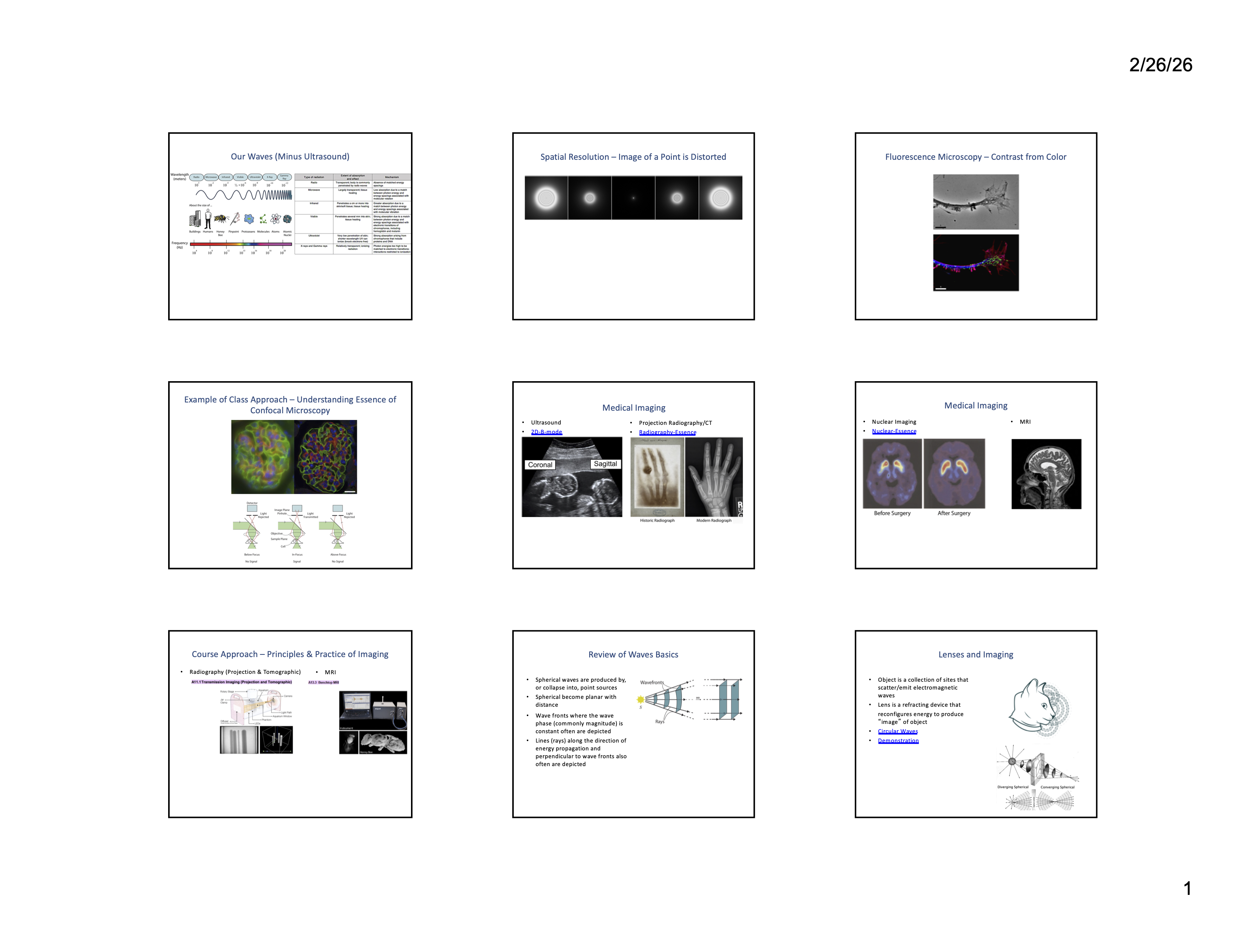
PowerPoint Slides
PowerPoint slides used by the instructor. Lectures were accompanied by a combination of PowerPoint slides, copious blackboard notes, demonstrations, simulations, and in-class activities.
 SP26.jpg)
In-Class Activities
In-class activities completed by students and discussed by the instructor approximately every other lecture. They were collected, but not graded, with the number completed contributing to student grades.
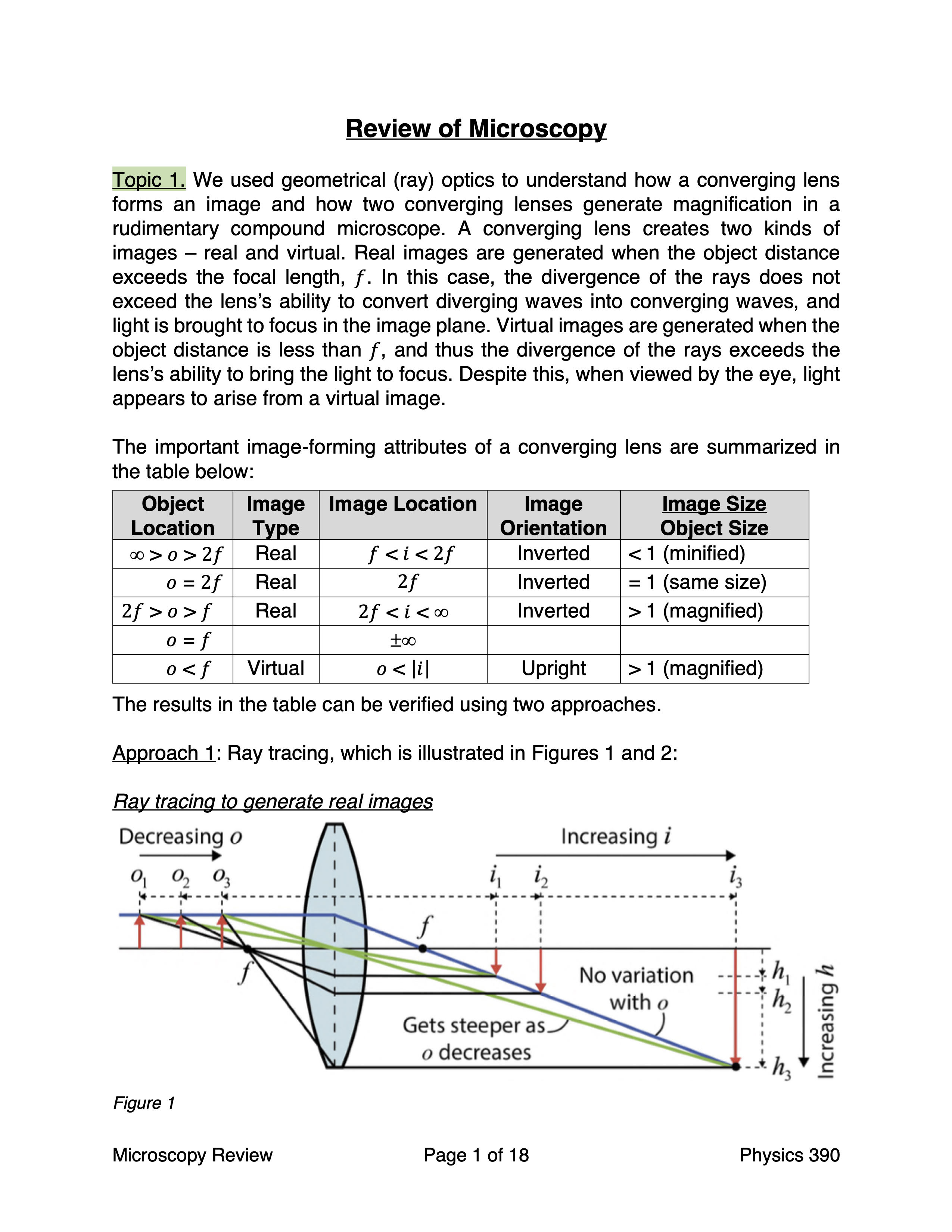
Course Reviews
Subject-matter reviews provided to students at the end of units on (1) microscopy, (2) medical imaging, and (3) image processing and analysis.
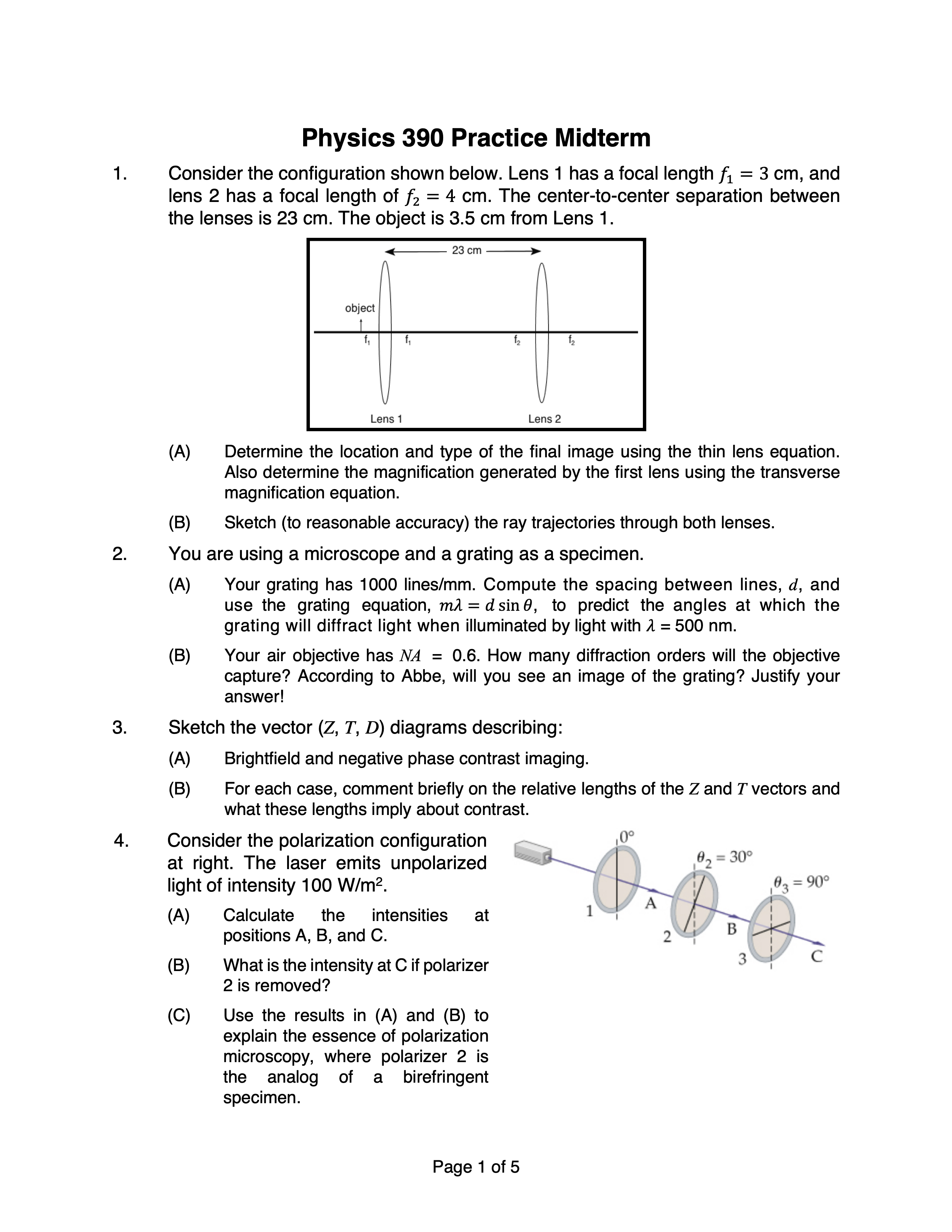
Practice Examinations
Practice examinations provided to students before midterms and the final examination.
Textbook Materials
This section includes a download link to images and other materials needed to complete selected Homework Problems in the book, a reference to a Homework Solution Manual, and a short list of errata.
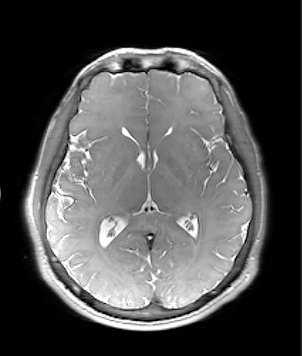
Homework Materials
Images and other materials needed to complete selected Homework Problems in the textbook, especially Chapters 9 (Image Processing) and 13 (Magnetic Resonance Imaging).
.jpg)
Homework Solution Manual
An 82-page Solution Manual showing worked solutions (not just answers) for every homework problem in the book is avaliable from the publisher to instructors adopting the book.